Fig. 5
- ID
- ZDB-FIG-250902-36
- Publication
- Rodriguez-Marquez et al., 2025 - Photodynamic Therapy Using a Heavy-Atom-Free G‑Quadruplex-Targeted Photosensitizer to Efficiently Regress Rhabdomyosarcoma Tumors In Vivo
- Other Figures
- All Figure Page
- Back to All Figure Page
|
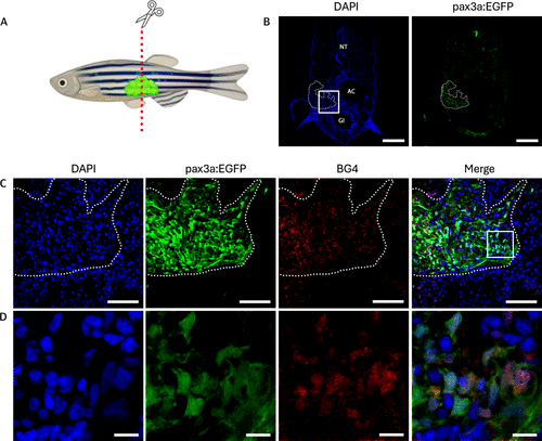
Elevated levels of G4 structures in RMS tissue sections of pax3a:EGFP zebrafish. (A) Schematic illustration of 2 month-old pax3a:EGFP zebrafish with RMS tumor. (B) Tissue section of the tumor, showing DAPI (left) and pax3a:EGFP (right). NT: neural tube, AC: abdominal cavity, GI: gastrointestinal tract. Dashed lines indicates where the tumor is located. Scale bar = 200 μm. (C) Zoom of the boxed region from (B), showing DAPI in blue, pax3a:EGFP expression in green, BG4 staining for G4 DNA in red, and a merged image of all channels. Scale bar = 50 μm. (D) Zoom of the boxed region from (C). Scale bar = 10 μm. |

