
Fig. 2
- ID
- ZDB-FIG-250902-33
- Publication
- Rodriguez-Marquez et al., 2025 - Photodynamic Therapy Using a Heavy-Atom-Free G‑Quadruplex-Targeted Photosensitizer to Efficiently Regress Rhabdomyosarcoma Tumors In Vivo
- Other Figures
- All Figure Page
- Back to All Figure Page
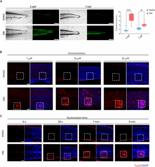
Uptake and activation of DBI in zebrafish. (A) Confocal laser scanning microscopy images of 2 days postfertilization (dpf) and 7 dpf AB zebrafish larvae treated with DMSO (control) or DBI, followed by illumination. Grayscale images (left) and corresponding green fluorescent signals (right) are shown. Scale bar = 200 μm. p < 0.01 (**), p < 0.0001 (****). (B,C) Apoptosis detection in zebrafish after DMSO or DBI treatment, followed by illumination, using different DBI concentrations: 1,10 and 25 μM (B) and different light exposure durations: 0 s to 2 min (C). Dashed squares indicate the illuminated areas. Apoptotic cells were stained with TUNEL (red), and nuclei were counterstained with DAPI (blue). Scale bar = 50 μm. |