Fig. 3
- ID
- ZDB-FIG-250902-34
- Publication
- Rodriguez-Marquez et al., 2025 - Photodynamic Therapy Using a Heavy-Atom-Free G‑Quadruplex-Targeted Photosensitizer to Efficiently Regress Rhabdomyosarcoma Tumors In Vivo
- Other Figures
- All Figure Page
- Back to All Figure Page
|
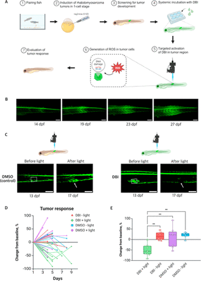
Effects of PDT on kRAS-induced RMS tumors in pax3a:EGFP zebrafish. (A) Schematic illustration of the induction of RMS tumors in zebrafish, followed by treatment with DBI and evaluation of the tumor response. (B) Representative image of RMS tumor progression in pax3a:EGFP zebrafish line, from 14 to 27 days post fertilization (dpf). The tumor region is identified by high levels of GFP expression. Scale bar = 200 μm. (C) Confocal images of tumors before and after treatment with DMSO + light (left) and DBI + light (right). Dashed squares indicate the illuminated areas. Tumor responses are indicated with the white arrows. Scale bar = 200 μm. (D) Comparison of tumor area, expressed as a percentage of initial size, after treatment across four conditions: DBI + light (treatment), and three controls: DBI – light, DMSO + light, and DMSO – light. (E) Percentage change in tumor area at 4–5 days post-treatment across the four conditions. p < 0.01 (**). |

