
Fig. 4 - Supplemental 1
- ID
- ZDB-FIG-250819-16
- Publication
- Chen et al., 2025 - Contraction-induced endocardial id2b plays a dual role in regulating myocardial contractility and valve formation
- Other Figures
- All Figure Page
- Back to All Figure Page
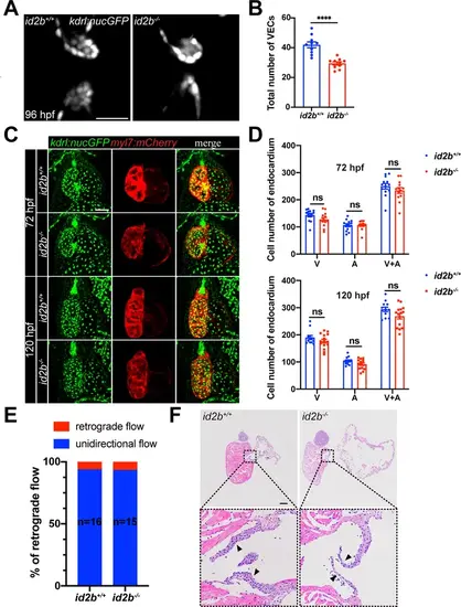
id2b-/- larvae exhibit a decreased number of valve endocardial cells while maintaining normal atrioventricular valve function. (A) Representative confocal images of valve endocardial cells in 96 hr post-fertilization (hpf) id2b+/+ and id2b-/- hearts carrying Tg(kdrl:nucGFP). (B) Quantification of the number of valve endocardial cells in id2b+/+ and id2b-/- hearts. VECs, valve endocardial cells. n=(10, 10). (C) Representative confocal images of 72 and 120 hpf id2b+/+ and id2b-/- hearts in Tg(kdrl:nucGFP); Tg(myl7:mCherry) transgenic background. (D) Statistical analysis of the number of endocardial cells in the ventricle (V), atrium (A), and combined (A+V) in id2b+/+ and id2b-/- hearts. n=(13, 13) (72 hpf); n=(12, 15) (120 hpf). (E) Quantification of blood flow patterns in 96 hpf id2b+/+ (n=16) and id2b-/- (n=15) hearts. (F) Hematoxylin and eosin (HE) staining of adult id2b-/- and id2b+/+ hearts after echocardiographic analysis in Figure 4F and G. Enlarged views of boxed areas are shown in the bottom panels. Data are presented as mean ± s.e.m. p-values were calculated by unpaired two-tailed Student’s t-tests. ****p<0.0001. ns, not significant. Scale bars, 20 μm (A), 50 μm (C), 200 μm (F). |