
Fig. 1
- ID
- ZDB-FIG-250819-11
- Publication
- Chen et al., 2025 - Contraction-induced endocardial id2b plays a dual role in regulating myocardial contractility and valve formation
- Other Figures
- All Figure Page
- Back to All Figure Page
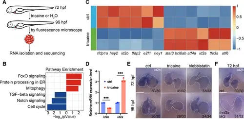
Identification of id2b as a blood flow-sensitive gene. (A) Schematic showing the experimental procedures, including treatment, heart collection, and RNA-sequencing of zebrafish embryonic hearts. (B) KEGG enrichment analysis depicting differentially expressed genes encoding transcription factors and transcriptional regulators between control (ctrl) and tricaine-treated embryonic hearts. Red and blue rectangles represent upregulated and downregulated gene sets, respectively. |log2fold change|≥0.585, adjusted p-value<0.1. Each replicate contains approximately 1000 hearts. (C) Heatmap exhibiting representative genes from KEGG pathways mentioned in (B). (D) Quantitative real-time PCR (qRT-PCR) analysis of id2b and id2a mRNA in control and tricaine-treated embryonic hearts. Data were normalized to the expression of actb1. Each sample contains ~1000 embryonic hearts. N=3 biological replicates. (E) In situ hybridization of id2b in 72 hr post-fertilization (hpf) and 96 hpf ctrl, tricaine (1 mg/mL), and blebbistatin (10 μM)-treated embryos. Numbers at the bottom of each panel indicate the ratio of representative staining. (F) In situ hybridization showing reduced id2b expression in tnnt2a morpholino-injected embryos at 72 hpf compared to control. Data are presented as mean ± s.e.m. Unpaired two-tailed Student’s t-tests were used to determine statistical significance. ***p<0.001. Scale bars, 50 μm. |