
Fig. 5 - Supplemental 2
- ID
- ZDB-FIG-250819-19
- Publication
- Chen et al., 2025 - Contraction-induced endocardial id2b plays a dual role in regulating myocardial contractility and valve formation
- Other Figures
- All Figure Page
- Back to All Figure Page
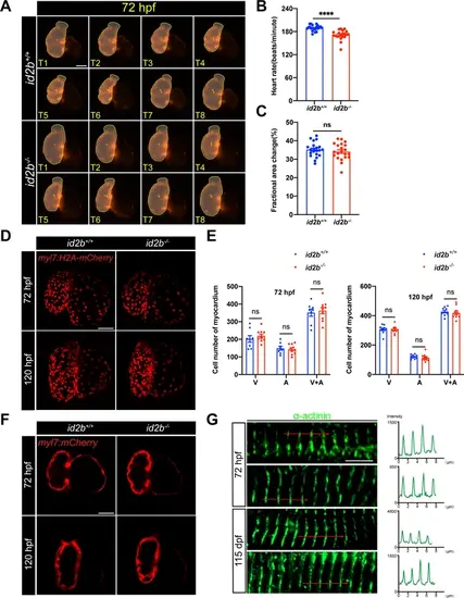
id2b-/- hearts develop normal trabeculae and sarcomeres. (A) Time-lapse imaging (from T1 to T8) illustrates the cardiac contraction-relaxation cycle of 72 hr post-fertilization (hpf) id2b+/+ and id2b-/- hearts carrying myl7:mCherry. (B and C) Heart rate and fractional area change in id2b-/- (n=21) and id2b+/+ (n=21) at 72 hpf. (D) Representative confocal z-stack of 72 and 120 hpf id2b+/+ and id2b-/- hearts with Tg(myl7:H2A-mCherry) transgene. (E) Quantification of the number of cardiomyocytes in the ventricle (V), atrium (A), and combined (A+V) in (D). n=(8, 10) (72 hpf); n=(10, 10) (120 hpf). (F) Representative confocal images of 72 and 120 hpf id2b+/+ and id2b-/- hearts carrying Tg(myl7:mCherry). (G) Confocal immunofluorescence images of α-actinin (green) in embryonic (72 hpf) and adult (115 dpf) hearts (left). Right: fluorescence intensity profiles for α-actinin. Data are presented as mean ± s.e.m. p-values were calculated by unpaired two-tailed Student’s t-tests. ****p<0.0001. ns, not significant. Scale bars, 50 μm (D and F), 5 μm (G). |