
FIGURE
Fig. 5
- ID
- ZDB-FIG-250814-57
- Publication
- Zou et al., 2025 - An effective fluorescent sensor for lipopolysaccharide-induced H2S detection and imaging in inflammatory cells, zebrafish, and mouse blood samples
- Other Figures
- All Figure Page
- Back to All Figure Page
Fig. 5
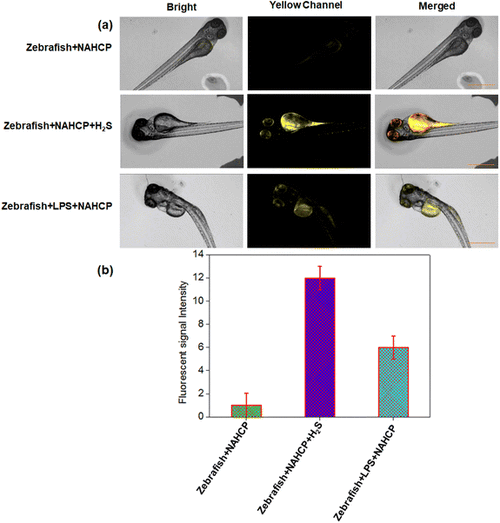
(a) Fluorescence inverted microscope imaging of NAHCP (10 μM) in living zebrafish. Top row: zebrafish was incubated with NAHCP (10 μM) for 30 min as the control group; middle row: zebrafish was incubated with NAHCP (10 μM) for 30 min and then with H2S 50 μM for 30 min; bottom row: zebrafish was incubated with LPS (1.0 μg mL−1) for 2 h and then with NAHCP (10 μM) for 30 min. (b) The quantitative analysis of the average fluorescence in the top row, middle row, and bottom row. λex = 420 nm, λem = (520–550) nm, scale bar: 10 μm, n = 3. |
Expression Data
Expression Detail
Antibody Labeling
Phenotype Data
Phenotype Detail
Acknowledgments
This image is the copyrighted work of the attributed author or publisher, and
ZFIN has permission only to display this image to its users.
Additional permissions should be obtained from the applicable author or publisher of the image.
Full text @ RSC Adv.