|
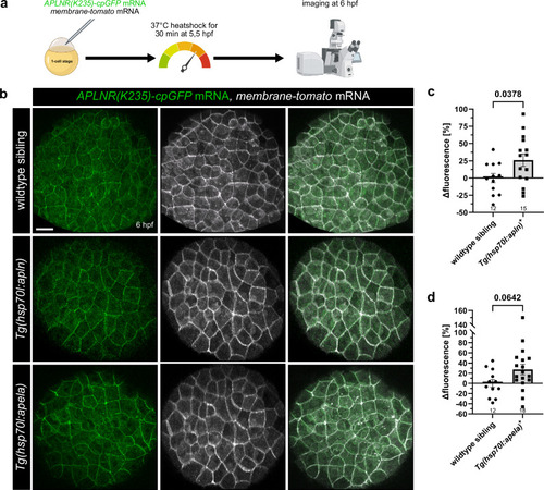
Ubiquitous Apelin and Apela overexpression activate APLNR(K235)-cpGFP biosensor in vivo. a Schematic illustration of the procedure. APLNR(K235)-cpGFP and membrane-tomato mRNA were injected into 1-cell stage zebrafish embryos. At 5.5 hours post-fertilization (hpf), injected embryos were heat shocked for 30 min at 37 °C to induce ubiquitous ligand overexpression and subsequently imaged at 6 hpf. b Representative confocal projection images of an injected wildtype sibling and transgenic Tg(hsp70l:apln) and Tg(hsp70l:apela) embryos at 6 hpf, respectively. Quantification of APLNR(K235)-cpGFP delta fluorescence intensity of wildtype siblings compared to embryos ubiquitously overexpressing the apln (c) and the apela (d) ligand. Each dot represents the mean of 10 measured cells per embryo. Data are presented as mean values ± SEM. (N number of embryos, n number of cells: c wildtype siblings N/n: 12/120, Tg(hsp70l:apln)+N/n: 15/150; d wildtype siblings N/n: 12/120, Tg(hsp70l:apela)+N/n: 18/180). Statistical analysis was performed by using a two-tailed unpaired Student’s t-test with Welch’s correction. Scale bars 30 µm. cpGFP circularly permuted GFP. Source data are provided as a Source Data file. a Created in BioRender. Schihada (2025) https://BioRender.com/vqh3y8e.
|