
Fig. 2
- ID
- ZDB-FIG-250709-23
- Publication
- Wieloch et al., 2025 - In vitro embryolethality testing during the peri-implantation stage using 3D mouse embryoids: comparison with 2D mouse cell cultures and the zebrafish embryo model
- Other Figures
- All Figure Page
- Back to All Figure Page
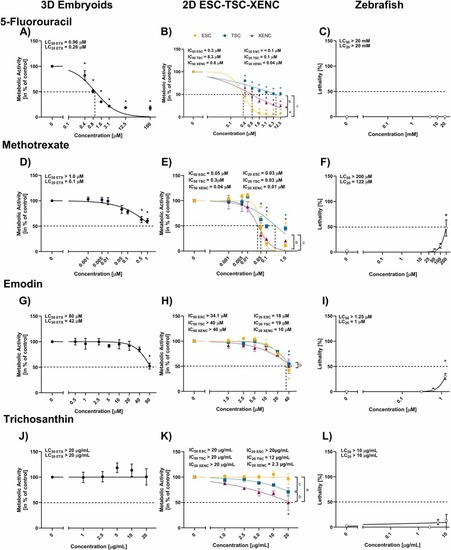
Metabolic activity of 3D embryoids (DIV4), 2D ESC-TSC-XENC and lethality of zebrafish embryos in the presence of 5-fluorouracil, methotrexate, emodin or trichosanthin. (A, D, G, J) Viability of DIV4 embryoids exposed for 48 h to increasing concentrations of 5-fluorouracil, methotrexate, emodin and trichosanthin by means of metabolic activity measured by CellTiterGlo-3D™ assay. (B, E, H, K) viability of ESC (yellow circles), TSC (blue squares) and XENC (magenta triangles) exposed for 48 h to increasing concentrations of 5-fluorouracil, methotrexate, emodin and trichosanthin by means of metabolic activity measured by CellTiterGlo-3D™ assay. (C, F, I, L) lethality of zebrafish embryos exposed for 48 h to 5-fluorouracil, methotrexate, emodin or trichosanthin, assessed according to the criteria described in TG 236 [29]. Results representing the mean ± SEM of at least three independent experiments. Statistical analysis of mouse embryoids and zebrafish embryos results was performed with one-way ANOVA followed by Dunnett post hoc comparison test, while for 2D cell cultures a two-way ANOVA followed by Tukey’s post hoc comparison test was performed to allow for evaluation of significant differences compared to the respective solvent control (*) and significant differences between cell lines: (a) ESC vs. XENC, (b) XENC vs. TSC (c) ESC vs. TSC. Significance threshold was established at p < 0.05 in all cases. |
Reprinted from Reproductive toxicology (Elmsford, N.Y.), , Wieloch, J., Blanco, J., Zordick, C., Ohnesorge, N., Schneider, M.R., Barenys, M., Knöspel, F., In vitro embryolethality testing during the peri-implantation stage using 3D mouse embryoids: comparison with 2D mouse cell cultures and the zebrafish embryo model, 108941108941, Copyright (2025) with permission from Elsevier. Full text @ Reprod. Toxicol.