FIGURE

Figure 5

ID
ZDB-FIG-250701-42
Publication
Rosa et al., 2025 - Evaluating TnP as a Potential Therapeutic Agent for Retinopathy in Zebrafish Models
Other Figures
All Figure Page
Back to All Figure Page
Figure 5

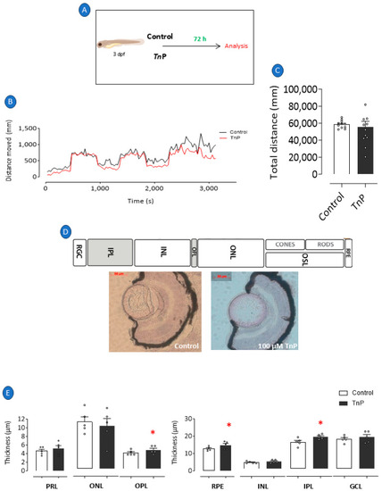
TnP’s per se effects. We prophylactically treated 3 dpf larvae with 100 µM TnP for 72 h (A). Larvae maintained at 28 °C in E2 0.5x medium only and under natural light were considered negative controls. Immediately after experimental period, all groups of larvae were evaluated for their visual–motor response (B) and distance traveled (C) and were subsequently killed and processed for histological analysis and H&E staining (D). Retinal layer thickness of zebrafish larvae (E). * p < 0.05 compared to control group. Bars represent mean ± SEM. Dots represent individual data points. RGL: retinal ganglion layer; IPL: inner plexiform layer; INL: inner nuclear layer; OPL: outer plexiform layer; ONL: outer plexiform layer; OSL: outer segment layer; RPE: retinal pigment epithelium; PRL: photoreceptor layer; GCL: ganglion cell layer.

Expression Data

Expression Detail
Antibody Labeling
Phenotype Data

Phenotype Detail
Acknowledgments
This image is the copyrighted work of the attributed author or publisher, and ZFIN has permission only to display this image to its users. Additional permissions should be obtained from the applicable author or publisher of the image. Full text @ Pharmaceuticals (Basel)