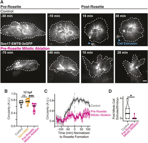
Pre-rosette mitotic events play an indispensable role in cell packing and subsequent cell extrusion during lumen formation. (A) Stills from a time-lapse movie of microtubule organization during KV formation (Sox17:EMTB-3xGFP; gray). KVs are outlined (white dashed line) for control (no ablation, top) and pre-rosette mitotic cell ablation conditions (bottom). Time is normalized to rosette formation (0 min); pre-rosette 0 to −70 min, post rosette is 0 to 30 min. Extruded cells are indicated with blue arrowheads and KV outline is highlighted by a white dashed line. Scale bar: 10 μm. See also Movie 4. (B) Truncated violin plot of KV circularity at 12 hpf. Control, non-mitotic cell ablation, and pre-rosette mitotic ablation conditions are shown. Median denoted with black line and quartile denoted by dashed line. n≥6 embryos were used across each condition. ***P<0.001 (one-way ANOVA with Dunnett's multiple comparison to non-mitotic cell ablation); ns, not significant. (C) Circularity of KV was measured over time normalized to rosette formation (marked by gray dashed line). Control (no ablation, gray) and ablation of pre-rosette mitotic events (four cells ablated, magenta) are shown. n≥6 embryos per condition. Error bars represent s.e.m. (D) Truncated violin plots of post mitotic cell extrusion events per embryo for control (no ablation) and ablation of pre-rosette mitotic events. Median denoted with black line and quartile denoted by dashed line. n≥6 embryos per condition. *P<0.05 (unpaired, two-tailed Student's t-test).
|
