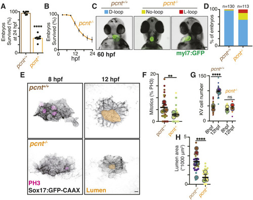
KV developmental defects and loss of mitotic events in pericentrin-null embryos are associated with reduced survival and increased left-right axis defects. (A) Percentage embryo survival from wild-type control crosses and embryos from pericentrin-null (pcnt−/−) crosses. n≥7 clutches shown at 24 hpf; each dot represents one clutch. Data are mean±s.e.m. ****P<0.0001 (unpaired, two-tailed Student's t-test). (B) Percentage pcnt−/− embryo survival from 1 hpf to 24 hpf across three clutches. Error bars represent s.e.m. (C) Representative images of 60 hpf wild-type and pcnt−/− fish with Tg(myl7:GFP) hearts shown with a normal cardiac loop (D-loop), an inverted loop (L-loop) and no loop. Scale bar: 100 μm. (D) Percentage of embryos with D-loop, L-loop and no loop calculated from wild-type and pcnt−/− clutches combined. n=2 clutches measured for wild type, n=4 clutches for pcnt−/− embryos; total embryo number ≥113 per condition. (E) Confocal projections of pre-lumen (8 hpf, left) and post-lumen (12 hpf, right) KVs lacking pericentrin (pcnt−/−) compared with wild-type control (pcnt+/+). KV cell membranes are marked with Sox17:GFP-CAAX (inverted gray), mitotic events with PH3 (magenta). Lumens were traced and highlighted in gold. Scale bar: 10 μm. (F-H) Percentage of mitotic KV cells (F), KV cell number (G) and lumen area (H) were measured for n≥23 embryos (small dots represent embryos color coded to match clutch) across n≥3 clutches (large dots represent clutch) per condition. Graphs are super plots (as defined by Lord et al., 2020) with mean of clutches shown ±s.e.m. **P<0.01, ****P<0.0001 (unpaired, two-tailed Student's t-test); ns, not significant.
|
