
Fig. 3
- ID
- ZDB-FIG-250529-8
- Publication
- Matsumata et al., 2025 - The habenula-interpeduncular nucleus-median raphe pathway regulates the outcome of social dominance conflicts in mice
- Other Figures
- All Figure Page
- Back to All Figure Page
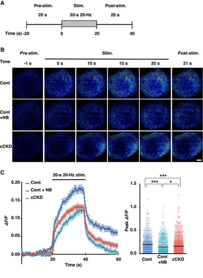
Impairment of ACh transmission in the vMHb-IPN pathway caused a reduction of the neural activities in the rIPN evoked by tetanic stimulation of the left fr (A) Time schedule of the electrical stimulation of fr. (B) Calcium imaging of brain slices of Cont and cCKD mice. NB, nAChR blockers. Scale bar, 100 μm. (C) Calcium signals in the rIPN of Cont (blue; n = 508 cells from 4 slices), Cont with nAChR blocker cocktails (Cont + NB) (pale blue; n = 607 cells from 6 slices), and cCKD (red; n = 1,000 cells from 9 slices) mice during tetanic stimulation (left). The traces are averaged for each condition. Error bars, mean ± SEM. Right panel represents the peak signal intensity during stimulation. Each dot indicates the peak ΔF/F of each cell of Cont, Cont + NB, and cCKD slices (H(2) = 49.14, p < 0.0001, Kruskal-Wallis test with Dunn’s multiple comparison test). Bars in (C), mean. Statistical significance was defined as ∗p < 0.05 and ∗∗∗p < 0.001. |