
Fig. 4
- ID
- ZDB-FIG-250522-10
- Publication
- Qin et al., 2025 - Age-dependent glial heterogeneity and traumatic injury responses in a vertebrate brain structure
- Other Figures
- All Figure Page
- Back to All Figure Page
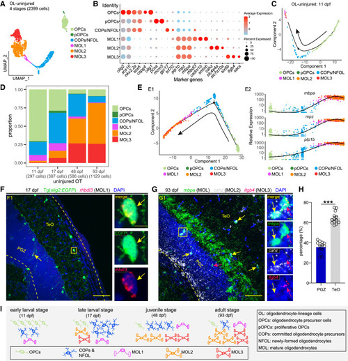
Temporal heterogeneity of oligodendrocyte-lineage cells under the physiological condition (A) UMAP plot showing re-clustering of oligodendrocyte-lineage cells (OL) from the uninjured OT across stages. Cells are colored by their identities. (B) Dot plot showing the expression of marker genes of each annotated cell type/subtype in (A). (C) Pseudo-time trajectory of OL in 11-dpf uninjured OT. (D) Histograms showing the proportion of each OL type/subtype at each stage. (E) Pseudo-time trajectories of OL from the uninjured OT across stages (E1) and the expression of myelin proteins along the trajectories (E2). (F) rhbdl3 in situ hybridization (red) combined with EGFP immunostaining (green) on the midbrain section of Tg(olig2:EGFP) at the late-larval stage (17 dpf). Yellow arrows indicate the rhbdl3+EGFP+ MOL1. (G) RNAscope of mbpa (green), cahz (white), and itgb4 (red) on the midbrain section of zebrafish at the adult stage (93 dpf). Yellow arrows indicate the cahz+mbpa+ MOL2, while red arrows indicate the itgb4+mbpa+ MOL3. (H) Relative ratio of cahz+mbpa+ MOL2 located in the TeO or PGZ of OT at 93 dpf in (G). (n = 13 sections/4 animals; mean ± SEM; parametric paired t tests; ∗∗∗p < 0.001). (I) Schematic illustration of oligodendrocyte-lineage cellular composition in uninjured OT across stages. In (F) and (G), dashed lines indicate OT boundaries. Boxed areas are shown magnified in the right panels. Scale bars, 50 μm (F1 and G1) and 5 μm (zoom-in images). Nuclei are visualized by DAPI (blue). |