
Fig. 3
- ID
- ZDB-FIG-250506-3
- Publication
- Wang et al., 2024 - Reprogramming miR-146b-snphb Signaling Activates Axonal Mitochondrial Transport in the Zebrafish M-cell and Facilitates Axon Regeneration After Injury
- Other Figures
- All Figure Page
- Back to All Figure Page
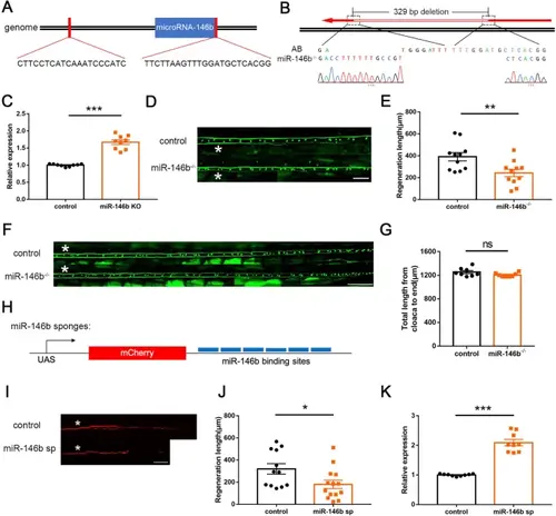
miR-146b loss-of-function inhibits M-cell axon regeneration by upregulating snphb. A Schematic of sgRNA target sites on both sides of microRNA-146b. B Sequencing results showed a ~329 bp deletion containing microRNA-146b in the miR-146b-/- mutants. C Quantitative RT-PCR assays showed a significant increase in snphb expression in the brains of 5-month-old adult miR-146b-/- mutants. D, E Representative images of control (T056) and miR-146b-/- M-cell axon regeneration results. Statistical results showed that miR-146b deletion inhibits axon regeneration in zebrafish M-cells. White asterisk denotes the injury site. Control: 390.9 ± 38.05 μm; miR-146b-/-: 242.1 ± 33.37 μm. Scale bar, 50 μm, ** P < 0.01, error bars indicate SEM. F, G No significant difference in the length of axons from the cloaca to the end between control (T056) and miR-146b-/- M-cell. A white asterisk denotes the defined sites. Control: 1253 ± 25.02 μm; miR-146b-/-: 1204 ± 11.70 μm. Scale bar, 100 μm, error bars indicate SEM. H Schematic diagram of miR-146b sponges (miR-146b sp). I, J Representative images of control (UAS-mCherry) and miR-146b sp axon regeneration results. Statistical results showed that miR-146b sp impeded axon regeneration in zebrafish M-cells. Control: 319.9 ± 46.98 μm; miR-146b sp: 180.0 ± 37.50 μm. White asterisk denotes the injury site. Scale bar, 50 μm, * P < 0.05, error bars indicate SEM. K Quantitative RT-PCR showed that the expression of snphb was significantly increased at 10 hpf in embryos expressing miR-146b sp. |