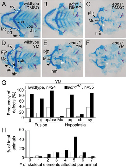
YM increases the prevalence and severity of lower jaw defects in edn1+/− larvae relative to wild-type larvae. (A-F) Representative flat-mounts of the viscerocranium from wild-type (A,D), edn1+/− (B,E) and edn1−/− (C,F) larvae treated with DMSO (A-C) or 100 μM YM (D-F) from 16 to 36 hpf. In A,B,D, white outlines highlight the symplectic cartilage (sy). In C,E,F, white asterisks indicate absence of the ceratohyal (ch). In D,E, black arrowheads indicate fusion of the jaw joint (jj). In D, the white arrowhead indicates fusion of the hyomandibular joint (hj). All skeletal preparations are 6 dpf. (G) Frequency of defects in seven Edn1/Ednra-dependent skeletal elements in YM-treated wild-type or edn1+/− larvae. Absence of structure, hypoplasia or fusion were scored as defects (ignoring sidedness). The pharmacogenetic interaction between edn1 and YM was determined to be statistically significant with a chi-square test (P=0.0001), comparing the number of defects in YM-treated edn1+/+ and edn1+/− larvae. (H) Percentage of individual larvae presenting with defects in the seven Edn1/Ednra-dependent structures. Individual larvae were scored for total number of skeletal elements affected (ignoring sidedness). Bar graphs are color-coded the same as G. All wild-type, edn1+/− or edn1−/− embryos treated with DMSO or YM are siblings from the same clutch. Scale bar: 100 μm. bsr, branchiostegal ray; hm, hyomandibular; Mc, Meckel's cartilage; op, opercle; pq, palatoquadrate; ptp, pterygoid process of the palatoquadrate.
|
