
Fig. 4
- ID
- ZDB-FIG-250324-39
- Publication
- Kobar et al., 2024 - tp53 R217H and R242H mutant zebrafish exhibit dysfunctional p53 hallmarks and recapitulate Li-Fraumeni syndrome phenotypes
- Other Figures
- All Figure Page
- Back to All Figure Page
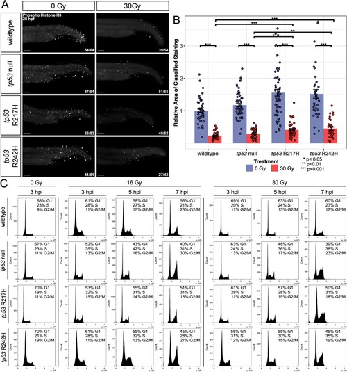
tp53 mutant embryos display increased proliferating cells and defective cell cycle checkpoints following IR compared to wildtype. (A) Immunofluorescence for pH3 performed on 28 hpf (3 hpi) embryos ±30 Gy. Representative images shown for each group as described by ratio in bottom right corner of each image. n = 3 replicates. (B) Bar graphs showing quantification of the relative area of pH3 positive cells in the tail region as determined using an ilastik Cell Profiler pixel classifier. The number of embryos used for analysis in each group is listed in (A). Statistical significance was determined by a two-way ANOVA and a Tukey's Honest Significant Difference test as shown by stars. Error bars represent standard error. (C) Flow cytometry histograms of DNA content in each cell cycle phase (G1, S, G2/M) at 3, 5, and 7 hpi dosed with 0, 16 or 30 Gy in wildtype, tp53 null, R217H/R217H, and R242H/R242H embryos. Embryos were enzymatically dissociated, fixed, permeabilized, and stained with FxCycle violet. Each plot was gated to 25,000 cells (±100 cells). Results and G1, S, and G2/M phase percentages are representative of three independent experiments. Scale bars, 100 μm. |
Reprinted from Biochimica et biophysica acta. Molecular basis of disease, 1871, Kobar, K., Tuzi, L., Fiene, J.A., Burnley, E., Galpin, K.J.C., Midgen, C., Laverty, B., Subasri, V., Wen, T.T., Hirst, M., Moksa, M., Carles, A., Cao, Q., Shlien, A., Malkin, D., Prykhozhij, S.V., Berman, J.N., tp53 R217H and R242H mutant zebrafish exhibit dysfunctional p53 hallmarks and recapitulate Li-Fraumeni syndrome phenotypes, 167612, Copyright (2024) with permission from Elsevier. Full text @ BBA Molecular Basis of Disease