FIGURE

Fig 6

ID
ZDB-FIG-250321-14
Publication
Nonarath et al., 2025 - The USH3A causative gene clarin1 functions in Müller glia to maintain retinal photoreceptors
Other Figures
All Figure Page
Back to All Figure Page
Fig 6

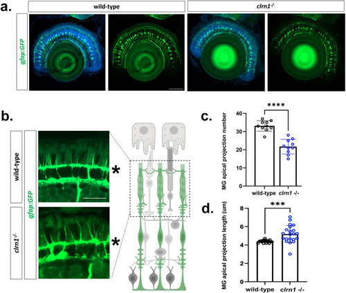
Actin is disorganized in the outer retina of clrn1-/- mutant zebrafish.

Transverse sections from 7 dpf wild-type and clrn1-/- Tg(gfap:GFP) zebrafish (a) showing the ordered spacing and radial expansion of Müller glia of both wild-type and clrn1 mutant retina. (b) Higher magnification images of Müller glia apical projections of wild-type and clrn1-/- zebrafish highlights disorganization of the mutant apical microvilli (c). Quantification of total Müller glia (MG) apical microvilli within a 40 µm2 region of the central retina of wildtype and clrn1-/- zebrafish (d). Müller glia (MG) apical microvilli length beyond the OLM (labeled with asterisk in b.) for wild-type and clrn1-/- 7 dpf zebrafish. (***p<0.005,****p<0.001; Students T-test). Scale bars = (a) 100 µm; (b) 50 µm. Data points in (c,d) represent measurements from n = 10 zebrafish each condition. For (d) data points are an average per zebrafish in which 9-22 projections quantified. Schematic in (b) created in BioRender under License: https://BioRender.com/x12n534.

Expression Data

Expression Detail
Antibody Labeling
Phenotype Data

Phenotype Detail
Acknowledgments
This image is the copyrighted work of the attributed author or publisher, and ZFIN has permission only to display this image to its users. Additional permissions should be obtained from the applicable author or publisher of the image. Full text @ PLoS Genet.