|
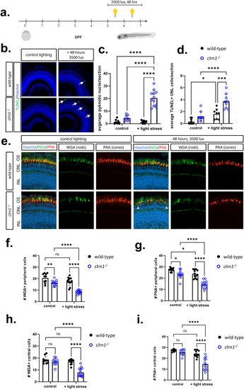
High-intensity light induces rod and cone cell death in clrn1-/- zebrafish. (a) Overview of light stress assay: larvae were treated with 3500 lux for 48 hours from 5-7 dpf. (b) Representative images of TUNEL-positive photoreceptors (white arrows) on cryosectioned retina from 7 dpf wild-type (upper panels) and clrn1-/- (lower panels) zebrafish revealed a significant increase in the (c) average number of pyknotic nuclei and (d) TUNEL-positive cells per section in the ONL of clrn1-/- zebrafish. (e) Retinal sections from 7dpf control or light-stressed wild-type (upper panels) and clrn1-/- zebrafish (lower panels) stained with wheat germ agglutinin (WGA, rods, green) and peanut agglutinin (PNA, cones, red) as well as Hoechst to label nuclei (white arrow heads denote pyknotic cells). Quantification of (f) WGA- and (g) PNA-positive outer segments along a 100 µm region of the peripheral retina revealed a significant decrease in rod and cone outer segments in control clrn1-/- zebrafish, which was exacerbated under light stress. Quantification of (h) WGA- and (i) PNA-positive outer segments along a 100 µm section of the central retina revealed a significant decrease in rod and cone outer segments in light-damaged clrn1-/- zebrafish. (*p<0.05, **p<0.01, ****p<0.001; Two-way ANOVA). Error bars=SD. Scale Bar = 100 µm. n=10-15 per genotype for each condition. OS, Outer Segment; ONL, Outer Nuclear Layer; INL, Inner Nuclear Layer. Schematic in (a) created in BioRender under License: https://BioRender.com/k07v149.
|