|
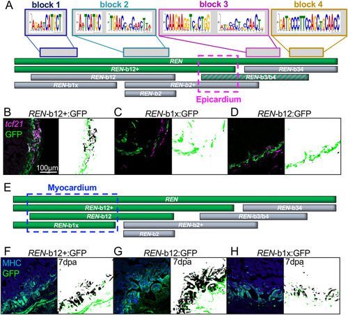
Myocardial REN-directed gene expression is separable from other cell types. (A) Schematic of full-length REN (top green bar) and the seven smaller REN fragments used in transgenic reporters. (B-D) GFP-positive fragments in epicardium (green) and GFP-negative (black). The epicardial-specific region is outlined in a pink dashed box in A. Left panels in B-D show hearts from the one positive REN-b12+ transgenic (B) and the two negative REN-b12 (C) and REN-b1x (D) reporters. Right panels show MIPAR renditions of colocalized areas (black) with excess GFP (green). (E) Same schematic as in A except REN fragments colored green are based on expression in muscle (α-MHC specific). (F-H) Left panels show hearts from the three positive reporter lines REN-b12+ (F), REN-b12 (G) and REN-b1x (H). Right panels show MIPAR renditions of colocalized areas (black) with excess GFP (green).
|