IMAGE

Fig. 2.

ID
ZDB-IMAGE-250307-60
Source
Figures for Rao et al., 2025
Image
Figure Caption

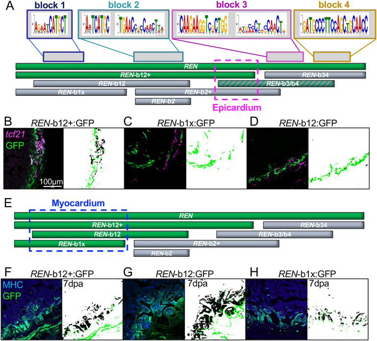
Fig. 2.

Myocardial REN-directed gene expression is separable from other cell types. (A) Schematic of full-length REN (top green bar) and the seven smaller REN fragments used in transgenic reporters. (B-D) GFP-positive fragments in epicardium (green) and GFP-negative (black). The epicardial-specific region is outlined in a pink dashed box in A. Left panels in B-D show hearts from the one positive REN-b12+ transgenic (B) and the two negative REN-b12 (C) and REN-b1x (D) reporters. Right panels show MIPAR renditions of colocalized areas (black) with excess GFP (green). (E) Same schematic as in A except REN fragments colored green are based on expression in muscle (α-MHC specific). (F-H) Left panels show hearts from the three positive reporter lines REN-b12+ (F), REN-b12 (G) and REN-b1x (H). Right panels show MIPAR renditions of colocalized areas (black) with excess GFP (green).

Acknowledgments
This image is the copyrighted work of the attributed author or publisher, and ZFIN has permission only to display this image to its users. Additional permissions should be obtained from the applicable author or publisher of the image. Full text @ Development