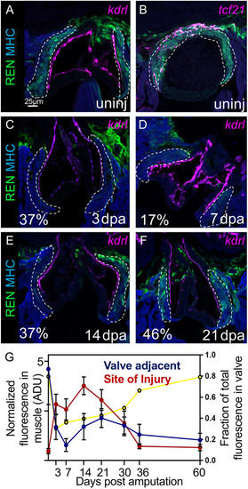
REN regulates gene expression in cardiac tissue surrounding valves in uninjured hearts. (A) Valves around the outflow tract in uninjured hearts are shown by endocardial reporter flk:Red (pink). REN:GFP is shown in green and muscle is stained with MHC (blue). (B) Both cardiac muscle (MHC, blue) and epicardium (tcf21 reporter, pink) colocalize with REN:GFP (green) around uninjured valves. (C-F) Same staining as in A, but this time on regenerating hearts at 3 dpa (C), 7 dpa (D), 14 dpa (E), and 21 dpa (F). Percentage of remaining GFP intensity is shown in the bottom left corner. (G) Mean GFP intensity in regions colocalizing with muscle (α-MHC). Cardiac valve regions are shown in blue, and site of injury is shown in red. The x-axis is a timeline for the days after amputation of the ventricle apex. The y-axis (left) is the normalized fluorescence and the y-axis (right) is the fraction of the total fluorescence that is found around valves (yellow line). The regions expected to be REN:GFP-positive in A-F are outlined with a white dashed line. However, quantitation was carried out for the entire muscle positive region. Data are mean±s.e.m.
|
