
Fig. 6
- ID
- ZDB-FIG-250305-34
- Publication
- Hundsdorfer et al., 2025 - ERK activation waves coordinate mechanical cell competition leading to collective elimination via extrusion of bacterially infected cells
- Other Figures
- All Figure Page
- Back to All Figure Page
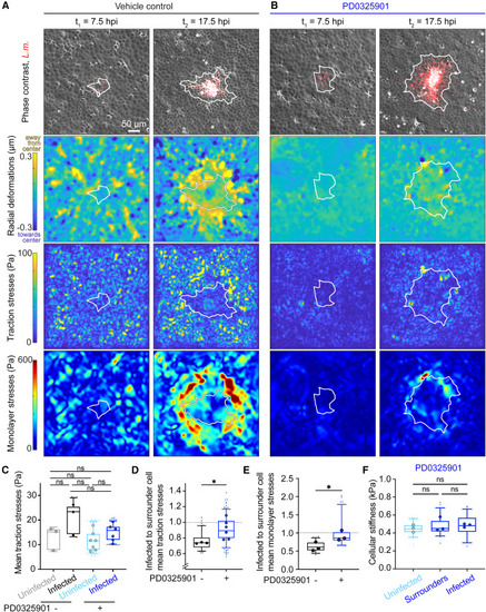
ERK inhibition stalls the increase in monolayer stresses in proximal surrounders and abolishes differences in stiffness between surrounders and infected cells (A and B) TFM and MSM on MDCK cells adherent on 3-kPa gels and infected at low MOI with L.m. such that single-infection foci emerge. Cells were treated with vehicle control (A) or 50 μM PD0325901 (B) at 4 hpi. Rows: overlay of phase contrast with L.m. fluorescence, radial deformations that cells impart on their matrix (μm), traction stresses (Pa), and monolayer stresses (Pa). Columns: time (hpi). White line: infection focus contour. (C) Box plots of mean traction stresses of L.m.-infected or not MDCK cells, treated or not with 50 μM PD0325901. ANOVA, ns: p > 0.05. (D and E) Box plots of ratio of traction stresses (D) or monolayer stresses (E) of infected to surrounder cells. WRST: ∗p < 0.05. For (C-E) N = 3 independent experiments were analyzed for cells followed from 10 to 16 hpi, dots show the mean of each sample. (F) Box plots of cell stiffness (kPa) at 24 hpi for uninfected, surrounder cells, and infected cells, for samples treated with 50 μM PD0325901 at 4 hpi. N = 58±3 cells were analyzed in each case originating from three independent experiments. ANOVA: ns: p > 0.05. See also Figure S6 and Video S5. |