Fig. 5
- ID
- ZDB-FIG-250305-33
- Publication
- Hundsdorfer et al., 2025 - ERK activation waves coordinate mechanical cell competition leading to collective elimination via extrusion of bacterially infected cells
- Other Figures
- All Figure Page
- Back to All Figure Page
|
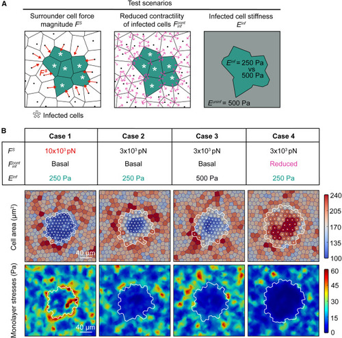
Modeling reveals that monolayer stress reinforcement of proximal surrounders is necessary for infected cell squeezing (A) Schematic of hybrid model. Infected cells are denoted by green asterisk. Proximal surrounders exert force Fs pointing toward the focus (left), infected cells versus surrounders can have varying contractility (middle) or varying cell stiffness (right). (B) Four cases were simulated (columns) by varying the parameters shown in (A). In each case, shown are the cell topology with cells color-coded by area (μm2, top), and monolayer stresses (Pa, bottom). (1) Infected cells contract, their stiffness is half compared to surrounders, Fs = 10 × 103 pN. (2) Infected cells contract, their stiffness is half compared to surrounders, Fs = 3 × 10³ pN. (3) Infected cells contract, their stiffness is the same as for surrounders, Fs = 3 × 10³ pN. (4) Infected cells contract less, their stiffness is half compared to surrounders, Fs = 3 × 10³ pN. White line: infection focus contour. Asterisks: infected cells. See also Figure S5 and Video S4. |

