
Fig. 2
- ID
- ZDB-FIG-250304-22
- Publication
- Sebo et al., 2025 - Activation of Wnt/β-catenin in neural progenitor cells regulates blood-brain barrier development and promotes neuroinflammation
- Other Figures
- All Figure Page
- Back to All Figure Page
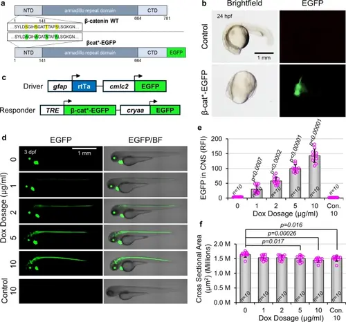
Generation of Dox-inducible transgenic lines to activate Wnt/β-catenin signaling in NPCs. (a) Schematic representation of endogenous β-catenin (β-catenin WT) and engineered constitutively active β-catenin fused to EGFP (β-cat*-EGFP). Serine and threonine residues S33, S37, T41, and S45 (yellow) were mutated to alanine (green), and EGFP was added in frame to the C terminus. (b) β-cat*-EGFP mRNA was synthesized and injected into single-cell embryos. At 24 hpf, injected embryos are dorsalized (no tail) and express EGFP. (c) Schematic of DNA constructs used to make the Tg(gfap:rtTA, cmlc2:EGFP) driver line and Tg(TRE:β-cat*-EGFP, cryaa:EGFP) responder line. Driver and responder lines are distinguished by green hearts (cmlc2:EGFP) or green lenses (cryaa:EGFP), respectively. (d) Representative lateral images of 3 dpf transgenic gfap;rtTA, TRE:β-cat*-EGFP larvae, or wild type siblings (Con.), treated with indicated Dox concentrations. (e) Quantification of EGFP fluorescence intensity within the CNS of larvae shown in (d) (n = 10 for all conditions). (f) Quantification of whole animal cross sectional area of larvae shown in (d) (n = 10 for all conditions). p-values were calculated for comparisons of all groups using the parametric one-way analysis of variance (ANOVA) with Tukey HSD post hoc test (no label = not significant). p-values directly above a bar indicate significant difference to all other groups by at least the level shown. Data are shown as mean ± standard deviation. |