|
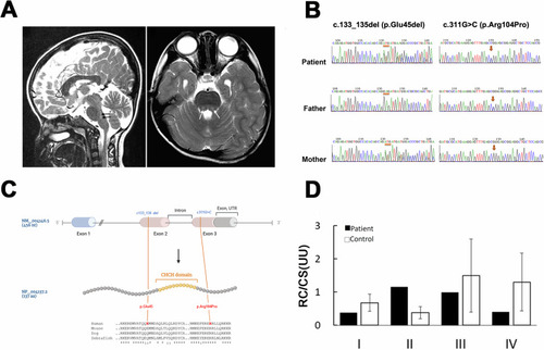
NDUFB7 is the disease causative gene. A The patient’s brain MRI T2-weighted sagittal (left) or axial image obtained at 4 years old reveals high-intensity pons lesions (gray and pointed by arrows). B Partial Sanger sequencing chromatographs of the patient and parents. The left panel shows the presence of a deletion (denoted by orange bars) in the patient and her mother, while the right panel shows a missense mutation (denoted by orange arrows) found in the patient and her father. C The upper panel shows the gene structure of the human NDUFB7 gene (NM_004146.5, 456 nucleotides (nt)), which has three exons and two introns (the intron between Exon 2 and 3 is indicated). The orange lines indicate two mutations in Exon 2 and 3 of the patient’s NDUFB7 gene. The NDUFB gene is translated to 137 amino acids (aa, NP_004137.2, a cartoon in the middle panel). A large portion of Exon 2 and 3 encodes a coiled-coil-helix-coiled-coil-helix (CHCH) domain marked in orange. The lower panel presents a sequence alignment of sequences comprising the mutations and the CHCH domains among human, mouse, dog, and zebrafish (“*” identical; “:” similar). D Respiratory chain (RC) activities in cultured fibroblasts from the patient expressed as a ratio (U/U) relative to citrate synthase (CS) activity. The patient’s cells exhibited higher Complex II activity but lower activities in other complexes.
|