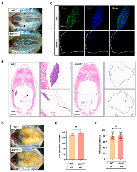
Dmrt1 regulates gonad development in males but not in females. (A) Anatomical observation of testes in WT and dmrt1−/− at 5 mpf. The black and blue arrowheads indicate the testes in WT and dmrt1−/− zebrafish, respectively. (B) Histological examination of testes in 5-month-old WT and dmrt1−/− zebrafish. Transverse section of the abdomen by HE staining. The black and blue arrowheads indicate the testes in WT and the severely regressed testicular tissue in dmrt1−/− zebrafish, while the blue dotted lines show the amplified images of testes in dmrt1−/− zebrafish. Scale bars, 1 mm. (C) Immunofluorescence staining of WT and dmrt1−/− testis transverse sections with Ddx4 antibody (green). The nuclei were stained by DAPI (blue). The white dotted lines show the testes in dmrt1−/− zebrafish. Scale bars, 50 μm. (D) Anatomical observation of ovary in 5-month-old WT and dmrt1−/− zebrafish. (E) Successful spawning rate of WT and dmrt1−/− females mating naturally with WT males at 5 mpf. There were three independent repetitions (n = 15). (F) Fertilization rate of WT and dmrt1−/− females by crossing with WT males at 5 mpf (n = 15). Data are represented as mean ± SD (ns, no significant).
|
