IMAGE

Figure 3

ID
ZDB-IMAGE-250228-74
Source
Figures for Ge et al., 2025
Image
Figure Caption

Figure 3

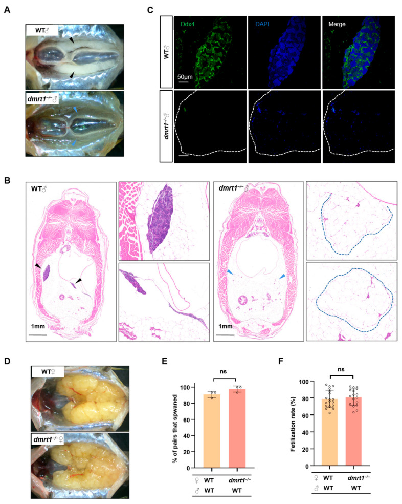
Dmrt1 regulates gonad development in males but not in females. (A) Anatomical observation of testes in WT and dmrt1−/− at 5 mpf. The black and blue arrowheads indicate the testes in WT and dmrt1−/− zebrafish, respectively. (B) Histological examination of testes in 5-month-old WT and dmrt1−/− zebrafish. Transverse section of the abdomen by HE staining. The black and blue arrowheads indicate the testes in WT and the severely regressed testicular tissue in dmrt1−/− zebrafish, while the blue dotted lines show the amplified images of testes in dmrt1−/− zebrafish. Scale bars, 1 mm. (C) Immunofluorescence staining of WT and dmrt1−/− testis transverse sections with Ddx4 antibody (green). The nuclei were stained by DAPI (blue). The white dotted lines show the testes in dmrt1−/− zebrafish. Scale bars, 50 μm. (D) Anatomical observation of ovary in 5-month-old WT and dmrt1−/− zebrafish. (E) Successful spawning rate of WT and dmrt1−/− females mating naturally with WT males at 5 mpf. There were three independent repetitions (n = 15). (F) Fertilization rate of WT and dmrt1−/− females by crossing with WT males at 5 mpf (n = 15). Data are represented as mean ± SD (ns, no significant).

Acknowledgments
This image is the copyrighted work of the attributed author or publisher, and ZFIN has permission only to display this image to its users. Additional permissions should be obtained from the applicable author or publisher of the image. Full text @ Int. J. Mol. Sci.