|
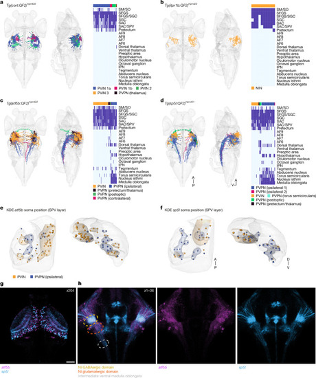
Neurons of a specific t-type may assume distinct morphologies and projection patterns. a, Sparsely labelled cort neurons were registered into the standard brain (n = 62). Right, anatomical matrix. AF, arborization field; IPN, interpeduncular nucleus; SAC, stratum album centrale; SGC, stratum griseum centrale; SM, stratum marginale; SO, stratum opticum. b, Sparsely labelled itpr1b neurons were registered into the standard brain (n = 10). Right, anatomical matrix. NIN, neuropil interneurons. c, Sparsely labelled atf5b neurons were registered into the standard brain (n = 77). Neurons are colour coded according to their m-type. Right, anatomical matrix. d, Sparsely labelled sp5l+ neurons were registered into the standard brain (n = 53). Right, anatomical matrix. A, anterior; P, posterior; V, ventral. e, atf5b interneurons (n = 49) and ipsilateral neurons (n = 18) were mirrored to the left hemisphere, and a KDE was measured according to their soma position, revealing separation of m-types along the anterior–posterior and dorsal–ventral axes. Transverse and coronal views of the SPV layer are shown. f, sp5l interneurons (n = 16) and ipsilateral neurons (n = 29) were mirrored to the left hemisphere, and a KDE was measured according to their soma position, revealing separation of m-types along the anterior–posterior and dorsal–ventral axes. g, Registered confocal stack of atf5b and sp5l transgenic fish. A single fish example of each transgenic line out of three specimens imaged. A single focal plane spanning the OT is shown. Scale bar, 50 μm. h, z-projection of the nucleus isthmi (NI) area, showing atf5b and sp5l projections forming collaterals at different parts of the nucleus isthmi.
|