
Fig. 3
- ID
- ZDB-FIG-250203-42
- Publication
- Shi et al., 2025 - An animal model recapitulates human hepatic diseases associated with GATA6 mutations
- Other Figures
- All Figure Page
- Back to All Figure Page
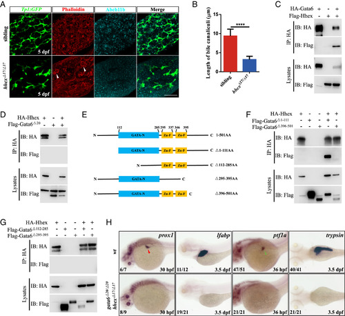
Gata6 interacts with Hhex physically and genetically. (A) Immuno- and dye staining of livers in the siblings (n = 6) and hhex mutants (n = 6) at 5 dpf. White arrowhead, hepatic cysts. GFP (green), intrahepatic bile duct cells; Phalloidin (red) and Abcb11b (cyan), bile canaliculi. (Scale bar, 50 μm.) (B) Quantification of the length of bile canaliculi in the figure (A). In the liver of hhex mutant, few residual bile canaliculi could be detected. (C and D) Coimmunoprecipitation assays of Hhex and Gata6 in HEK293T cells. (E) Schematic drawings of wild type and mutant Gata6 proteins. (F and G) Coimmunoprecipitation assays of Hhex and mutated variants of Gata6. (H) WISH results of wild type and gata6−/−; hhex−/− double mutants at 30 hpf and 3.5 dpf using prox1 (hepatoblasts), lfabp (hepatocytes), ptf1a, and trypsin probes. Red arrowhead, liver bud; Green arrowhead, exocrine pancreas. Data are mean ± SD, ****P < 0.0001, two-tailed unpaired t test. |