
- Title
-
An animal model recapitulates human hepatic diseases associated with GATA6 mutations
- Authors
- Shi, W., Yi, X., Ruan, H., Wang, D., Wu, D., Jiang, P., Luo, L., Ma, X., Jiang, F., Li, C., Wu, W., Luo, L., Li, L., Wang, G., Qiu, J., Huang, H.
- Source
- Full text @ Proc. Natl. Acad. Sci. USA
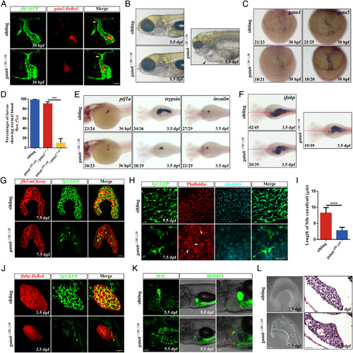
The knockout of gata6 causes various defects in zebrafish. (A) Tg(flk1:GFP) and Tg(gata1:DsRed) were used to label endocardium (green) and erythrocyte (red), respectively (n = 9 siblings, n = 10 gata6 mutants). Yellow arrowhead, the presumed outflow tract. (Scale bar, 50 μm.) (B) Brightfield views of the siblings and gata6 mutants at 5.5 dpf. Black arrowhead, pericardial edemas. (C) Analysis of gata4 and gata5 expression in the heart tube at 30 hpf. (D) Percentages of embryos with normal blood flow at 3 dpf (n = 328 siblings, n = 82 gata6Δ20/Δ20; gata5+/+, n = 108 gata6Δ20/Δ20; gata5+/Δ4). (E) WISH showing ptf1a (exocrine pancreatic progenitors), trypsin (exocrine pancreas), and insulin (β cell) expression at the indicated stages. (F) Expression of ifabp (intestinal epithelium) at 3.5 dpf. Red dashed lines, the midline. (G) Tg(flk1:mCherry) and Tg(Tp1:GFP) were used to reveal vascular network (red) and intrahepatic biliary network (green), respectively (n = 6 siblings, n = 4 gata6 mutants). (Scale bar, 50 μm.) (H) Immuno- and dye staining of livers in the siblings (n = 6) and gata6 mutants (n = 7) at 7.5 dpf. GFP (green), intrahepatic bile duct cells; Phalloidin (red), F-actin cytoskeleton enriched in bile canaliculi; Abcb11b (cyan), a bile salt export pump located in bile canaliculi. Phalloidin staining showed hepatic cysts (white arrowhead) in gata6 mutant livers. (Scale bar, 50 μm.) (I) Quantification of the length of bile canaliculi in the figure (H). Few abnormal bile canaliculi were detected in the gata6 mutant liver. (J) Confocal projection images showing the expression of Tg(lfabp:DsRed) and Tg(Tp1:GFP) in the liver at 3.5 dpf (n = 12 siblings, n = 9 gata6 mutants). (Scale bar, 50 μm.) (K) Confocal images of 2F11 antibody staining (n = 5 siblings, n = 5 gata6 mutants) or BODIPY-FL C5:0 feeding (n = 7 siblings, n = 11 gata6 mutants). Red asterisk, the gallbladder; Red arrowhead, BODIPY droplets. (Scale bar, 50 μm.) (L) Confocal projection images showing the morphology of the siblings (n = 5) and gata6 mutants (n = 5) livers. Green asterisk, hepatic cysts. (Scale bar, 50 μm.) Hematoxylin-eosin staining of the liver sections in the siblings (n = 4) and gata6 mutants (n = 4) at 7.5 dpf. Red asterisk, the cystic spaces. (Scale bar, 20 μm.) Data are mean ± SD, ***P < 0.001, ****P < 0.0001, two-tailed unpaired t test. |
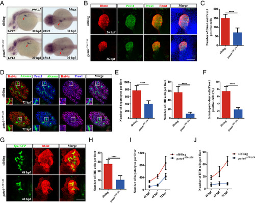
The loss of gata6 in zebrafish confers hepatobiliary abnormalities. (A) WISH with markers of hepatoblast (prox1 and hhex) at 30 hpf. Red arrowhead, liver bud. (B) Confocal images of Bhmt and Prox1 antibody staining (n = 14 siblings, n = 16 gata6 mutants) in the liver bud at 36 hpf. (Scale bar, 50 μm.) (C) Quantification of the Bhmt and Prox1 positive cells in the figure (B). (D) Confocal single-plane images of the liver immunostained for Hnf4α (red), Alcama (green), and Prox1 (blue) in siblings (n = 8) and gata6 mutants (n = 9) at 3 dpf. Alcama+ (high levels); Prox1+; Hnf4α−, IHD cells; Hnf4α+; Prox1+, hepatocytes. (Scale bar, 50 μm.) (E) Number of hepatocytes and IHD cells in siblings (n = 7) and gata6 mutants (n = 9). (F) The percentages of IHD cells in the Prox1 positive cells. (G) Immunostaining of Tg(Tp1:GFP) embryos at 48 hpf using Bhmt antibody (n = 17 siblings, n = 15 gata6 mutants). (Scale bar, 50 μm.) (H) Number of IHD cells in siblings and gata6 mutants in figure (G). (I and J) Quantification of the number of hepatocytes and IHD cells in siblings and gata6 mutants from 48 hpf to 72 hpf. Data are mean ± SD, ****P < 0.0001, two-tailed unpaired t test. |
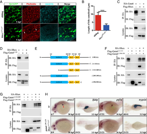
Gata6 interacts with Hhex physically and genetically. (A) Immuno- and dye staining of livers in the siblings (n = 6) and hhex mutants (n = 6) at 5 dpf. White arrowhead, hepatic cysts. GFP (green), intrahepatic bile duct cells; Phalloidin (red) and Abcb11b (cyan), bile canaliculi. (Scale bar, 50 μm.) (B) Quantification of the length of bile canaliculi in the figure (A). In the liver of hhex mutant, few residual bile canaliculi could be detected. (C and D) Coimmunoprecipitation assays of Hhex and Gata6 in HEK293T cells. (E) Schematic drawings of wild type and mutant Gata6 proteins. (F and G) Coimmunoprecipitation assays of Hhex and mutated variants of Gata6. (H) WISH results of wild type and gata6−/−; hhex−/− double mutants at 30 hpf and 3.5 dpf using prox1 (hepatoblasts), lfabp (hepatocytes), ptf1a, and trypsin probes. Red arrowhead, liver bud; Green arrowhead, exocrine pancreas. Data are mean ± SD, ****P < 0.0001, two-tailed unpaired t test. |

Gata6 and Hhex synergistically activates lrh-1 transcription. (A) Immuno- and dye staining of livers in the siblings (n = 6) and lrh-1 mutants (n = 6) at 7.5 dpf. 2F11 (green), intrahepatic bile duct cells; Phalloidin (red) and Mdr1 (cyan), bile canaliculi. (Scale bar, 50 μm.) (B) Quantification of the length of bile canaliculi in the figure (A). Few bile canaliculi could be observed in the lrh-1 mutant liver. (C) The lrh-1 expression revealed by WISH in gata6 and hhex mutants at 30 hpf. Red arrowhead, liver bud. (D) An illustration of the −3 kb promoter region of zebrafish lrh-1 gene highlighted with nine putative GATA binding sites (black square). The putative GATA binding sites were predicted on the website https://jaspar.genereg.net/. (E) Luciferase assays of the zebrafish −3 kb lrh-1 promoter in HepG2 cells. (F) Luciferase assays of the zebrafish −1.5 kb and −3 kb lrh-1 promoters with Gata6 in HepG2 cells. (G) ChIP-qPCR analysis of Gata6 occupancy on the lrh-1 promoter in Tg(pIDM:HA-gata6) larvae. As per the platform https://jaspar.genereg.net/, the three binding sites (A, B, and C in D) with the higher scores were selected. The coding region of lrh-1 in exon 7 was used as a negative control. (H) ChIP-seq reads across lrh-1 promoter loci with Tg(pIDM:HA-gata6) larvae upon DOX treatment. Gata6 bound to −1.6 kb of promoter loci of lrh-1 in zebrafish. (I) Luciferase assays of the zebrafish −3 kb lrh-1 promoter with Hhex and wild type and mutated variants Gata6 in HepG2 cells. (J) The expression of prox1 in gata6−/−; hhex−/− double mutants with the overexpression of lrh-1 by Tg(hsp70:HAM-lrh-1) heat-shocked at 14 hpf. Red arrowhead, liver bud. Data are mean ± SD, **P < 0.01, ***P < 0.001, ****P < 0.0001, ns, nonsignificant, two-tailed unpaired t test. |
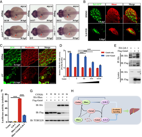
The coordinated interactions among Gata6, Hhex, and Lrh-1 modulate the output of β-catenin signaling. (A) WISH showing myca expression at 30 hpf. Red arrowhead, liver bud. (B) Immunostaining of Tg(Tp1:GFP) embryos with or without XAV939 treatment (18 hpf-54 hpf) at 3.5 dpf using Bhmt antibody (red, hepatocytes) (n = 10/10, DMSO, n = 11/14 XAV939). (Scale bar, 50 μm.) (C) Immuno- and dye staining of livers in the siblings (n = 14) and prt mutants (n = 12) at 5.5 dpf. (Scale bar, 50 μm.) (D) TOP-FLASH assays wherein Lrh-1 and the increasing amounts of Gata6 expression in HEK293T cells. (E) Coimmunoprecipitation assays of Gata6 and Lrh-1 in HEK293T cells. (F) Luciferase assays of the lrh-1 promoter with Gata6, Hhex, and Lrh-1 in HepG2 cells. (G) Immunoassay of Flag-Gata6 and HA-Hhex with or without CHX treatment at the indicated time points in HEK293T cells. (H) A model to depict the molecular mechanism underlying liver development in zebrafish. Data are mean ± SD, ****P < 0.0001, two-tailed unpaired t test. |
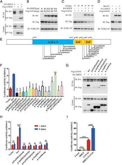
GATA6 mutant variants exhibit anomalous interaction with HHEX. (A) Coimmunoprecipitation assays of GATA6 and HHEX in HEK293T cells. (B–D) Analysis of the effect of HHEX on GATA6 protein stability. (E) Schematic drawing showing the positions of 15 mutated GATA6 variants associated with hepatobiliary malformations. GATA-N, transcriptional activation domain (light blue); ZnF, zinc finger domain (deep yellow). (F) Luciferase activity of a −3 kb human LRH-1 promoter with wild type and 15 mutated GATA6 variants in HepG2 cells. (G) Coimmunoprecipitation assays of HHEX and five representative mutated GATA6 variants. (H) Luciferase activity of a −3 kb human LRH-1 promoter with HHEX and five representative mutated GATA6 variants in HepG2 cells. (I) Luciferase assays of the −3 kb zebrafish lrh-1 promoter with human GATA6 and HHEX in HepG2 cells. Data are mean ± SD, *P < 0.05, **P < 0.01, ****P < 0.0001, ns, nonsignificant, two-tailed unpaired t test. |