
Fig. 2
- ID
- ZDB-FIG-250203-41
- Publication
- Shi et al., 2025 - An animal model recapitulates human hepatic diseases associated with GATA6 mutations
- Other Figures
- All Figure Page
- Back to All Figure Page
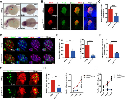
The loss of gata6 in zebrafish confers hepatobiliary abnormalities. (A) WISH with markers of hepatoblast (prox1 and hhex) at 30 hpf. Red arrowhead, liver bud. (B) Confocal images of Bhmt and Prox1 antibody staining (n = 14 siblings, n = 16 gata6 mutants) in the liver bud at 36 hpf. (Scale bar, 50 μm.) (C) Quantification of the Bhmt and Prox1 positive cells in the figure (B). (D) Confocal single-plane images of the liver immunostained for Hnf4α (red), Alcama (green), and Prox1 (blue) in siblings (n = 8) and gata6 mutants (n = 9) at 3 dpf. Alcama+ (high levels); Prox1+; Hnf4α−, IHD cells; Hnf4α+; Prox1+, hepatocytes. (Scale bar, 50 μm.) (E) Number of hepatocytes and IHD cells in siblings (n = 7) and gata6 mutants (n = 9). (F) The percentages of IHD cells in the Prox1 positive cells. (G) Immunostaining of Tg(Tp1:GFP) embryos at 48 hpf using Bhmt antibody (n = 17 siblings, n = 15 gata6 mutants). (Scale bar, 50 μm.) (H) Number of IHD cells in siblings and gata6 mutants in figure (G). (I and J) Quantification of the number of hepatocytes and IHD cells in siblings and gata6 mutants from 48 hpf to 72 hpf. Data are mean ± SD, ****P < 0.0001, two-tailed unpaired t test. |