
Fig. 1
- ID
- ZDB-FIG-250128-62
- Publication
- Revenu et al., 2024 - Myosin 1b regulates intestinal epithelial morphogenesis via interaction with UNC45A
- Other Figures
- All Figure Page
- Back to All Figure Page
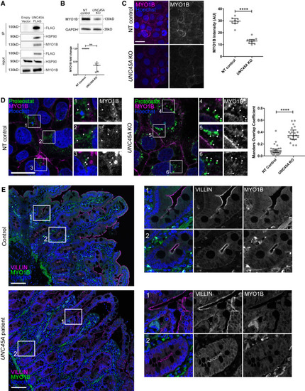
MYO1B interacts with UNC45A and is misfolded in UNC45A-depleted cells and in biopsies from UNC45A-deficient patient (A) Coimmunoprecipitation of UNC45A with MYO1b and HSP90 in Caco-2 cells transduced with empty vector (EV) or FLAG/MYC-UNC45A. (B) Western blot analysis of UNC45A Caco-2 KO cells and NT control lysates using anti-MYO1b antibody and relative quantification (n = 5). (C) Immunohistochemistry analyses and quantification of MYO1b intensity in NT control (n = 8) and UNC45A KO (n = 9) Caco-2 cells. Pictures are maximal projections of confocal stacks; Hoechst labels nuclei. Scale bar, 30 μm. (D) Confocal sections of NT control and UNC45A KO Caco-2 cells treated with the proteasome inhibitor MG132 and stained for MYO1b and with the aggresome probe Proteostat and quantification of the colocalization (NT n = 32; KO n = 21). Hoechst labels nuclei. Scale bar, 30 μm. Boxed areas shown in insets are enlarged 2×. Arrowheads point at Proteostat-labeled protein aggregates and highlight colocalization with MYO1B proteins in UNC45A KO cells. (E) Confocal sections of a human biopsy from a control and from a UNC45A LOF patient immunolabeled for the microvilli marker Villin and for MYO1b; Hoechst labels nuclei. Scale bar, 100 μm. Boxed areas shown in insets are enlarged 3× and highlight the apical localization of MYO1b in duodenal control tissue at the base of the villi (1) and in crypts (2). Data presented in (B), (C), and (D) are shown as median and interquartile range; Mann-Whitney test ∗∗p < 0.01, ∗∗∗∗p < 0.0001. |