FIGURE

Figure 1

ID
ZDB-FIG-250127-135
Publication
Ma et al., 2025 - A Lateral Line Specific Mucin Involved in Cupula Growth and Vibration Detection in Zebrafish
Other Figures
All Figure Page
Back to All Figure Page
Figure 1

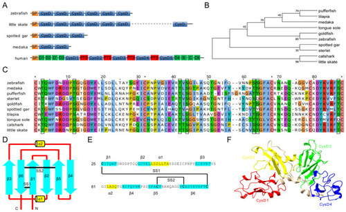
Structure, multiple alignment, and phylogenetic tree of Mucin-5AC proteins. (A) Schematic diagram of Mucin-5AC domains in zebrafish, little skate, spotted gar, medaka, and human. SP, signal peptide; CysD, cysteine rich domain; PTS, central proline, threonine, serine (PTS)-rich sequence. Green bars represent von Willebrand domains. (B) Phylogenetic tree of the first or second CysD domains of Mucin-5ACs. (C) Multiple alignment of the first or second CysD domain of Mucin-5ACs. Asterisks represent conserved cysteines (C). Black boxes indicate conserved serines (S) and threonines (T). (D) Secondary structure element of CysD1 in zebrafish Mucin-5AC. (E) Amino-acid sequence arrangement of CysD1 in zebrafish Mucin-5AC. The blue arrows (β1–β6) represent anti-parallel beta-strands and the yellow cylinders (α1–α2) represent short alpha helices. The black lines (SS1–SS2) mark disulfide bonds. (F) Three-dimensional structure of zebrafish Mucin-5AC (CysD1-CysD4) predicted by Alphafold.

Expression Data

Expression Detail
Antibody Labeling
Phenotype Data

Phenotype Detail
Acknowledgments
This image is the copyrighted work of the attributed author or publisher, and ZFIN has permission only to display this image to its users. Additional permissions should be obtained from the applicable author or publisher of the image. Full text @ Int. J. Mol. Sci.