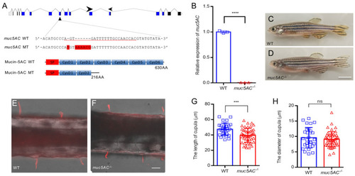
Mutation of muc5AC reduces the length of cupula in zebrafish larvae. (A) Schematic diagrams of muc5AC gene knockout. Top panel shows the scheme of muc5AC gene locus. Blue bars represent coding exon. Black bars represent 3′-or 5′-UTR. Arrowhead marks the location of gRNA. Arrows show the position of primers for qRT-PCR. Middle panel shows the sequence around gRNA target in wild-types and mutants. The gRNA target sequence is underlined. The insertion nucleotides are marked with red background. Bottom panel shows the truncated Mucin-5AC protein predicted according to the DNA insertion. (B) qRT-PCR analysis of muc5AC gene in muc5AC mutant and wild-type larvae at 5 dpf. (C,D) Representative images of wild-type and muc5AC mutant zebrafish at 3 months post-fertilization (mpf). Scale bar: 5 mm. (E,F) Representative images of cupulae in wild-type and mutant larvae at 5 dpf. Scale bar: 50 μm. (G) Quantification of cupula length in wild-type and muc5AC mutant larvae at 5 dpf. (H) Quantification of cupula diameter in wild-type and muc5AC mutant larvae at 5 dpf. ***, p < 0.001; ****, p < 0.0001; ns, not significant.
|
