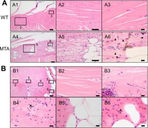
Adipose accumulation and degeneration observed in the skeletal muscle of the slc25a20 mutant. (A) Representative images of the skeletal muscle from hematoxylin and eosin (HE)-stained paraffin sections of the wild-type fish (A1–3) and the mutant (A4–6). (A1, A4) Low-magnification images of the skeletal muscles in the dorsal trunk. Squares correspond to the magnified regions shown in the following images. Scale bars, 100 μm. (A2, A5) Magnified images. Scale bars, 50 μm. (A3, A6) High-magnification images. The back arrowheads indicate eosin-negative membranes surrounding eosin-positive nuclei. Scale bars, 10 μm. (B) Representative images of the skeletal muscle in the tail region near the trunk from the HE-stained paraffin section of the mutant (the same section as in Fig. 3B). The body surface of the tail was sloped, so in the cut surface of this section, the tissue is closer to the skin from the left side to the right side. (B1) Low-magnification image. Squares correspond to the magnified regions shown in the following images. Scale bars, 100 μm. (B2–6) High-magnification images. Arrows indicate the faint traces of sarcomere structure. Scale bars, 10 μm.
|
