|
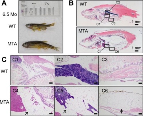
Adipose tissue accumulation inside and outside the coelomic cavity of the slc25a20 mutant. (A) Appearance of zebrafish used for tissue analysis. (B) Representative hematoxylin and eosin (HE) staining of 3-μm paraffin sections from the individuals showing a cross-section of the whole body. Squares correspond to the magnified regions shown in the following images. (C) Marked accumulation of adipose tissue in the mutant (C4–6) compared to the wild-type fish (C1–3). The images between the liver and the intestine (C1, C4), near the testes (C2, C4), and just outside of the coelomic cavity (C3, C6) are shown. Image C5 was taken from a different section than the one in B. The back arrows indicate abnormal accumulation of adipose tissue. Scale bar, 100 μm.
|