
Fig. 1
- ID
- ZDB-FIG-250114-20
- Publication
- Hishida et al., 2024 - Homozygous slc25a20 zebrafish mutant reveals insights into carnitine-acylcarnitine translocase deficiency pathogenesis
- Other Figures
- All Figure Page
- Back to All Figure Page
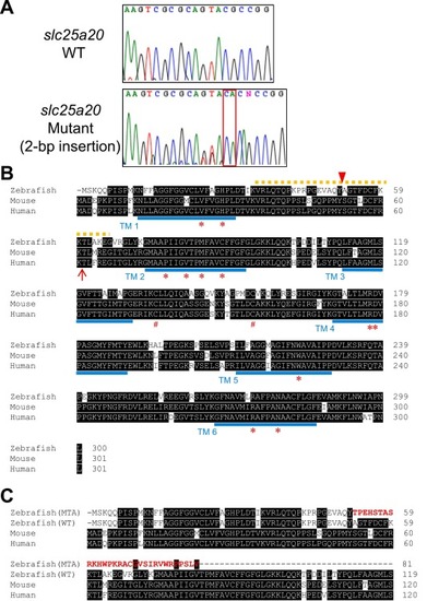
Generation of slc25a20 mutant zebrafish. (A) Sequences of wild-type (WT) and slc25a20 mutant fish are shown. The red square indicates the insertion with a 2-base pair addition. The N (G or A) after the insertion site represents a genetic polymorphism. (B) Protein sequence of zebrafish Slc25a20. Comparison of the amino acid sequence of SLC25A20 among D. rerio, M. musculus, and humans. Sequence alignment was conducted using Clustal Omega (https://www.ebi.ac.uk/jdispatcher/msa/clustalo) [58]. Shaded residues indicate conserved amino acids. The red arrowhead indicates the position of the insertion mutation in D. rerio, isolated in this study. The orange dotted line indicates the site encoded by exon 2 in zebrafish. Blue bars indicate the positions of the 6 presumed transmembrane domains [5]. Red asterisks and sharps indicate the amino acid residues involved in binding substrate molecules and in cell redox sensing and control, respectively [7,22]. The red arrow indicates the position of the frameshift mutation (p.Lys61fs) found in a severely ill patient [27,35]. (C) Protein sequence of zebrafish Slc25a20 (MTA) and its comparison of the amino acid sequence. The red bold characters indicate the unrelated 30-amino acid sequence after the frameshift. |