FIGURE

Figure 8

ID
ZDB-FIG-250110-89
Publication
Carra et al., 2024 - How Tumors Affect Hemodynamics: A Diffusion Study on the Zebrafish Transplantable Model of Medullary Thyroid Carcinoma by Selective Plane Illumination Microscopy
Other Figures
All Figure Page
Back to All Figure Page
Figure 8

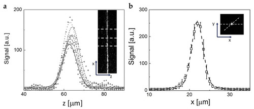
(a) Example of the profiles of the cross section of the light sheet image acquired by means of a reflecting mirror (640 × 512 pixels, 880 µm × 700 µm). The inset reports the image of the sheet cross-section on which the dashed lines indicate only a few of the sections that are analyzed in the main panel. The lines in the main panel are the best fit of the profiles to a Gaussian function with an average fitting FWHM = 7.8 ± 1.3 µm. (b) Example of the average cross-section of a single 1 μm fluorescent bead imaged in the light sheet used to determine the value of the PSF of the setup (640 × 512 pixels, 880 × 700 µm2). The dashed line is the best fit Gaussian function to the data with FWHM = 3.2±0.4 μm. The inset shows an example of the image of a microsphere in the light sheet.

Expression Data

Expression Detail
Antibody Labeling
Phenotype Data

Phenotype Detail
Acknowledgments
This image is the copyrighted work of the attributed author or publisher, and ZFIN has permission only to display this image to its users. Additional permissions should be obtained from the applicable author or publisher of the image. Full text @ Int. J. Mol. Sci.