
Fig. 4
- ID
- ZDB-FIG-250106-12
- Publication
- Dernovšek et al., 2024 - First dual inhibitors of human topoisomerase IIα and Hsp90 C-terminal domain inhibit the growth of Ewing sarcoma in vitro and in vivo
- Other Figures
- All Figure Page
- Back to All Figure Page
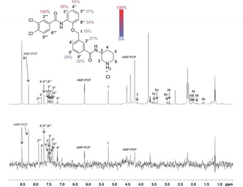
1D 1H STD NMR spectra for the compound 48 recorded at a Hsp90β:ligand ratio of 1:100 and 600 MHz. The structure of 48 shows the proton nomenclature and the color-coded relative degrees of saturation of the individual not-overlapped protons. The STD amplification factors were normalized to the intensity of the signal with the largest STD effect. The reference STD spectrum (top) with proton assignment and the difference STD spectrum (bottom) are shown. The unassigned proton signals between 3.5 and 3.8 ppm belong to protein buffer with glycerol. Signal for 3 partially overlaps with signals of AMP-PCP and signal for 2a partially overlaps with signals of protein buffer with glycerol. Intensities of 2, 3, 4, 5 and 6 are under detection limit in the difference STD spectrum. Since signals for 4′, 5‴ and 6‴ are overlapped, their STD amplification factors could not be determined. The proton signals were calibrated to the DSS signal at 0.0 ppm. The spectra are not to scale. |