
Fig. 6
- ID
- ZDB-FIG-241219-23
- Publication
- Lee et al., 2024 - CXCR3-CXCL11 Signaling Restricts Angiogenesis and Promotes Pericyte Recruitment
- Other Figures
- All Figure Page
- Back to All Figure Page
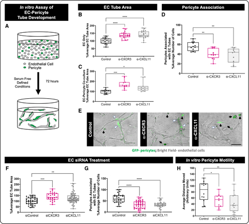
Endothelial cell (EC) CXCR3 (CXC motif chemokine receptor 3) and CXCL11 (CXC motif chemokine ligand 11) inhibition promotes EC tube formation and suppresses pericyte recruitment in vitro. A, Schematic of 3-dimensional (3D) collagen type I EC-pericyte tube assembly assays. ECs and pericytes are used in a 5:1 ratio, and the pericytes are GFP (green fluorescent protein) labeled for visualization. B and C, EC% average tube area measured from assays without (B; n=22) and with (C; n=12 control, n=11 α-CXCR3, and n=11 α-CXCL11, independent cultures) pericytes present. Cultures were treated with neutralizing antibodies to CXCR3 (α-CXCR3) or to CXCL11 (α-CXCL11), and cultures were then allowed to assemble tubes for 72 hours. Neutralization of CXCR3 or CXCL11 leads to expanded EC tube area regardless of the presence or absence of pericytes. D, When pericytes are present, neutralization of CXCR3 or CXCL11 leads to reduced pericyte association with EC tubes (n=12 control, n=12 α-CXCR3, and n=10 α-CXCL11, independent cultures). E, Representative image of the 3D EC-pericyte cocultures at 72 hours. ECs are shown by bright field, and pericytes are GFP labeled. White arrow heads indicate pericytes associated with EC tubes, and black arrows indicate pericytes that are not associated with EC tubes. F and G, To determine whether these effects are EC autonomous, we utilized siRNA to suppress CXCR3 (siCXCR3) or CXCL11 (siCXCL11) only in ECs, then incorporated these ECs into the 3D collagen gel assay with wild-type pericytes. EC tube area is increased in siCXCR3 and siCXCL11 conditions compared with the siControl condition (F; n=37 siControl, n=26 siCXCR3, and n=67 siCXCL11, independent cultures). Pericyte association with EC tubes is decreased following EC siCXCR3 or siCXCL11 treatment (G; n=29 siControl, n=37 siCXCR3, and n=30 siCXCL11, independent cultures). All data are normalized to the control condition. H, Quantification of the distance that pericytes migrate in 3D collagen gel assays treated with neutralizing antibodies suggests that pericyte motility is impaired when CXCR3-CXCL11 signaling is inhibited (n=12, independent cultures). For B through D and F through H, each dot represents an individual 3D collagen assay. Statistical analyses were performed using 1-way ANOVA with Dunnett multiple comparisons test (B–D, G, and H) and Kruskal-Wallis test with Dunn multiple comparisons (F). Omnibus ANOVA P values (before the post hoc tests) are <0.0001 (B), 0.0006 (C), 0.0021 (D), 0.0003 (F), <0.0001 (G), and 0.0081 (H). Data are presented as box plots that display the median value with first and third quartiles and min/max bars. *P<0.05, **P<0.005, ***P<0.0005, ****P<0.0001. |