FIGURE

Figure 2

ID
ZDB-FIG-241129-49
Publication
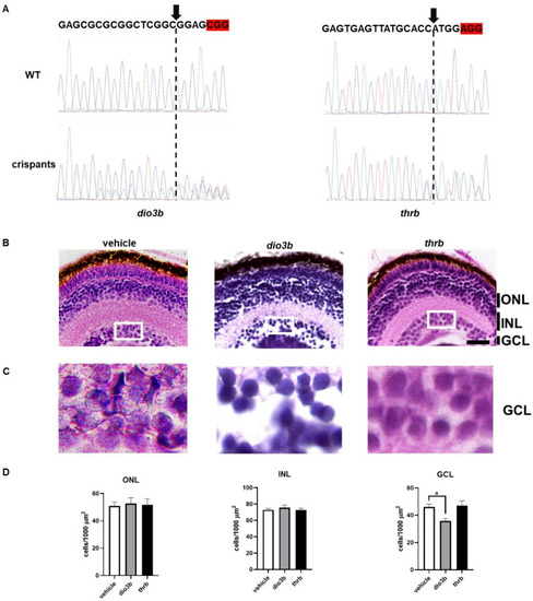
Lazcano et al., 2024 - Ontogeny of Thyroid Hormone Signaling in the Retina of Zebrafish: Effects of Thyroidal Status on Retinal Morphology, Cell Survival, and Color Preference
Other Figures
All Figure Page
Back to All Figure Page
Figure 2

Comparison of dio3b and thrb crispants at retinal level. (A) Electropherograms from a fragment of the PCR amplicon sequence for dio3b and thrb of the vehicle-microinjected controls and the corresponding crispants. At the top, the sgRNA sequence is denoted, and the PAM sequence is highlighted in red. In the crispants, a decrease in the resolution of the electropherogram is evident around the canonical cutting site indicated with an arrow and a dotted line. (B) Representative histological cross-sections of retinas of vehicle-injected controls and thrb and dio3 crispants (5–7 retinas per condition) stained with H&E. (C) Amplification of the GCL (insets in (B)). (D) Quantification of the number of cells/1000 µm2. ONL = outer nuclear layer, INL = inner nuclear layer, GCL = ganglion cell layer. Statistical analyses were performed using one-way ANOVA and Tukey post hoc tests. * = p < 0.05. Scale bar = 20 µm.

Expression Data

Expression Detail
Antibody Labeling
Phenotype Data
Fish:
Knockdown Reagents:
Observed In:
Stage: Day 5

Phenotype Detail
Acknowledgments
This image is the copyrighted work of the attributed author or publisher, and ZFIN has permission only to display this image to its users. Additional permissions should be obtained from the applicable author or publisher of the image. Full text @ Int. J. Mol. Sci.