
FIGURE
Fig. 8
Fig. 8
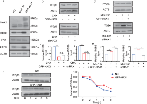
HAX1 enhances ITGB6 translation. (a) Western blot analysis of the protein levels in HUVECs-NC and HUVECs-shHAX1. (b, c) After 24 h treatment with CHX, cells were used for Western blot analysis (d, e) After 24 h treatment with MG132, cells were used for Western blot analysis. (f) HUVECs-NC and HUVECs-GFP-HAX1 were treated with CHX (100 µg/ml) and collected at different time points for Western blot analysis |
Expression Data
Expression Detail
Antibody Labeling
Phenotype Data
Phenotype Detail
Acknowledgments
This image is the copyrighted work of the attributed author or publisher, and
ZFIN has permission only to display this image to its users.
Additional permissions should be obtained from the applicable author or publisher of the image.
Full text @ J Extracell Vesicles