
Fig. 1
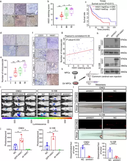
HAX1 overexpression is related to NPC metastasis and angiogenesis. (a) Representative immunohistochemical analysis (IHC) images of HAX1 staining in tumour tissues of different clinical grades from patients with NPC, bar: 100 µm. (b) Statistical comparison of HAX1 scores in different clinical grades. (*p < 0.05, **p < 0.01, one-way ANOVA). (c) Kaplan–Meier curves showing the correlation between HAX1 expression and overall survival by the log-rank test (Data from TCGA-HNSCC). (d) Tumours were analysed by IHC for microvessel densities (MVDs), bar: 100 µm. (e) Statistical comparison of number of MVDs in different clinical grades (**p < 0.01, ***p < 0.001, one-way ANOVA). (f) Representative IHC images of MVDs and HAX1 staining. (g) Spearman correlation between HAX1 score and MVDs in NPC. Pearson correlation coefficient (r2) and p-values are shown. (h) Up: the HAX1 levels of CNE2-NC, CNE2-shHAX1 and CNE2-GFP-HAX1 were measured by Western blotting. Down: The HAX1 levels of 6-10B-NC, 6-10B-shHAX1 and 6-10B-GFP-HAX1 were measured by Western blotting. (i) Visualisation of lung metastasis after intravenous injection of transfected NPC cells in BALB/c mice. (j) Mean fluorescence intensity (*p < 0.05, one-way ANOVA). (k) Schematic diagram of common cardinal vein injection in zebrafish. (l) NPC cells (red) were injected into the common cardinal vein of zebrafish at 48 hpf, and photographed using a confocal microscope (representative of three embryos per group, bar: 200 µm (m) Quantification of migratory cell numbers. (****p < 0.0001, Student's t-test) |