FIGURE

Figure 2.

ID
ZDB-FIG-241106-20
Publication
Haug et al., 2024 - Differential Localization and Functional Roles of mGluR6 Paralogs in Zebrafish Retina
Other Figures
All Figure Page
Back to All Figure Page
Figure 2.

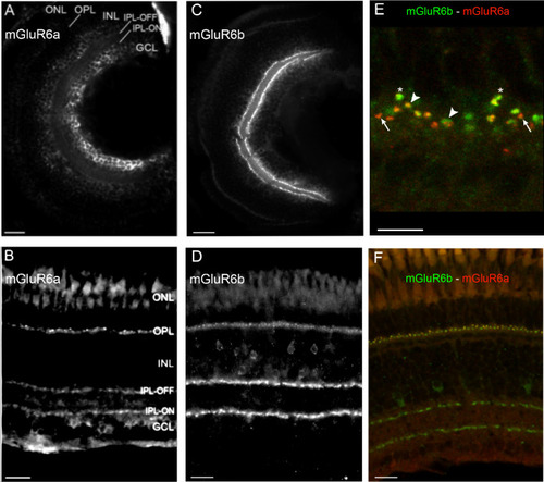
Retinal expression of mGluR6 paralogs in larval and adult zebrafish. (AD) Immunohistochemical labeling of mGluR6a on larval (A) and adult (B) and mGluR6b on larval (C) and adult (D) retinal cross sections. (E, F) Costaining of mGluR6a (red) and mGluR6b (green) on adult retinal cross sections. In (E), a zoom-in on the OPL shows where mGluR6b (asterisk) and mGluR6a (long arrow) do not overlap and where mGluR6a and mGluR6b expression overlaps (large arrowhead) (see Supplementary Table S2). GCL, ganglion cell layer; INL, inner nuclear layer; IPL‐OFF, off‐layer of the inner plexiform layer; IPL‐ON, on‐layer of the inner plexiform layer; ONL, outer nuclear layer; OPL, outer plexiform layer. Scale bars: 20 µm, except E, where the scale bar = 5 µm.

Expression Data
Genes:
Fish:
Anatomical Terms:
Stage Range: Day 5 to Adult

Expression Detail
Antibody Labeling
Phenotype Data

Phenotype Detail
Acknowledgments
This image is the copyrighted work of the attributed author or publisher, and ZFIN has permission only to display this image to its users. Additional permissions should be obtained from the applicable author or publisher of the image. Full text @ Invest. Ophthalmol. Vis. Sci.