FIGURE

Fig. 4

ID
ZDB-FIG-241031-17
Publication
Baird et al., 2024 - Rare homozygous cilia gene variants identified in consanguineous congenital heart disease patients
Other Figures
All Figure Page
Back to All Figure Page
Fig. 4

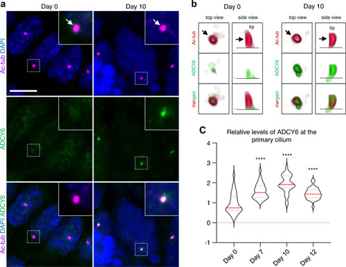
Temporal localization of ADCY6 to primary cilia during cardiomyogenesis. ADCY6 accumulate at primary cilia during differentiation of P19.CL6 cells into cardiomyocytes. a Representative images P19.CL6 cells at day 0 (stem cells) and day 10 (cardiomyocytes). Arrows point to primary cilia. Scale bar, 10 µm. b 3D visualization of representative cilia at day 0 and day 10 of differentiation. Arrow points to the primary cilium. Asterisk marks the ciliary base. c Violin plots of quantification of ADCY6 fluorescence levels at primary cilia during days 0, 7, 10 and 12 of differentiation. Asterisks indicate P values: ****P < 0.0001

Expression Data

Expression Detail
Antibody Labeling
Phenotype Data

Phenotype Detail
Acknowledgments
This image is the copyrighted work of the attributed author or publisher, and ZFIN has permission only to display this image to its users. Additional permissions should be obtained from the applicable author or publisher of the image. Full text @ Hum. Genet.