
Fig. 8
- ID
- ZDB-FIG-241022-56
- Publication
- Ding et al., 2024 - Inhibition of PNCK inflames tumor microenvironment and sensitizes head and neck squamous cell carcinoma to immune checkpoint inhibitors
- Other Figures
- All Figure Page
- Back to All Figure Page
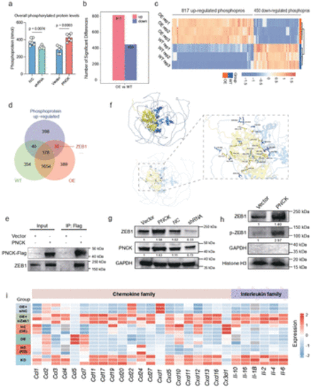
PNCK regulates ZEB1 nuclear translocation and reshapes the tumor immune-suppressive microenvironment. (a) Quantification of overall phosphorylated protein levels in various cell samples to assess changes in the phosphorylation status. A two-tailed unpaired Student’s t-test was performed. Data are presented as mean values±SD. (b) Differential phosphorylated proteins between the PNCK overexpression group and the WT group. (c) Heatmap showing the expression pattern of differentially phosphorylated proteins in the PNCK OE and WT groups. (d) Venn diagram showing the intersection of upregulated phosphorylated proteins and proteins interacting with PNCK in WT and OE groups. (e) Co-immunoprecipitation (CO-IP) validation demonstrates the interaction between PNCK and ZEB1. (f) Surface diagram of the docking model and their interfacing residues between ZEB1 and PNCK protein (ZEB1, blue; PNCK, yellow; hydrogen bond interaction, dotted line). (g) Western blot analysis shows changes in ZEB1 expression levels in response to alterations in PNCK expression. (h) Western blot analysis shows the upregulation of ZEB1 and phosphorylated ZEB1 (p-ZEB1) in the nuclear protein fraction of the PNCK OE group. GAPDH was used as a cytoplasmic protein loading control, and Histone H3 was used as a nuclear protein loading control. (i) Cytokine array analysis of numerous immune-related cytokines across multiple experimental groups, including KD, NC(KD), OE, NC(OE), OE+siZEB1, and OE+siNC. |