FIGURE

Fig. 5

ID
ZDB-FIG-241017-34
Publication
Armistead et al., 2024 - A sphingolipid rheostat controls apoptosis versus apical cell extrusion as alternative tumour-suppressive mechanisms
Other Figures
All Figure Page
Back to All Figure Page
Fig. 5

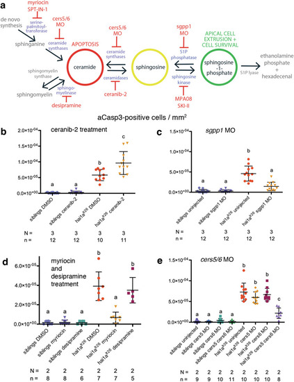
Manipulation of the sphingolipid rheostat enzymes or ceramide de novo synthesis alters cell death in the hai1afr26 mutant.

a Schematic of sphingolipid rheostat showing inhibition of ceramidases with ceranib-2, inhibition of sphingosine kinases with MPA08 or SKI-II, inhibition of S1P phosphatase with a MO targeting sgpp1, inhibition of serine palmitoyltransferase with myriocin or SPT-IN-1 and sphingomyelinases with desipramine, and MOs targeting cers5 and cers6 at the level of de novo synthesis and salvage from sphingosine. b Quantification of aCasp3-positive cells in the tail fins of 4 dpf embryos upon DMSO or ceranib-2 treatment, normalised to fin area. c Quantification of aCasp3-positive cells in the tail fins of control or sgpp1 MO-injected 4 dpf embryos, normalised to fin area. d Quantification of aCasp3-positive cells in the tail fins of 4 dpf embryos upon DMSO, myriocin, or desipramine treatment, normalised to fin area. e Quantification of aCasp3-positive cells in the tail fins of control or cers5 alone, cers6 alone, or both cers5 and cers6 MO-injected 4 dpf embryos, normalised to fin area. For all graphs, means were compared using one-way ANOVA with post-hoc Tukey’s multiple comparison test. N = number of biological replicates, n = number of fish per condition.

Expression Data

Expression Detail
Antibody Labeling
Phenotype Data

Phenotype Detail
Acknowledgments
This image is the copyrighted work of the attributed author or publisher, and ZFIN has permission only to display this image to its users. Additional permissions should be obtained from the applicable author or publisher of the image. Full text @ Cell Death Dis.