
Fig. 3
- ID
- ZDB-FIG-241016-3
- Publication
- McCann et al., 2024 - Emc1 is essential for vision and zebrafish photoreceptor outer segment morphogenesis
- Other Figures
- All Figure Page
- Back to All Figure Page
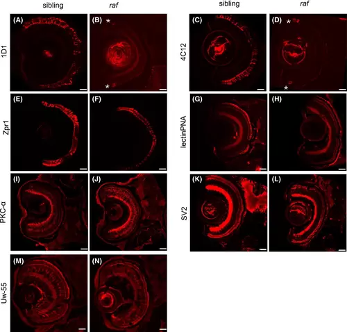
raf zebrafish have reduced outer retinal markers but inner retinal markers are not changed. (A–N): Images of retinal cross sections from wild-type siblings (A, C, E, G, I, K, and M) and raf−/− (B, D, F, H, J, L, and N) stained with antibodies to retinal cell populations (red); 1D1 (A and B), 4C12 (C and D), Zpr1 (E and F), lectin PNA (G and H), PKC-α (I and J), SV2 (K and L), and Uw-55 (M and N). Asterisks show faint 1D1/4C12 staining in the peripheral retina in raf−/− mutant images. Sections were generated with cryostat and imaged with a confocal microscope with 40x objective (A–N). Larvae were 6 dpf (A-L) and 4 dpf (M and N). Scale bar = 20 μm (A–N). |