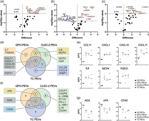
Quantitative analysis of the targets of the inflammation panel by PEA revealed distinctive receptor‐dependent tuning of the PEV proteins. PEVs isolated by iodixanol cushion ultracentrifugation were analysed for 80 inflammation panel targets. The GPVI (n = 4; biological replicates representing 16 donors), CLEC‐2 (n = 3; biological replicates representing 12 donors) and TC PEVs (n = 3; biological replicates representing 12 donors) were compared to the PEVs from unstimulated platelets (US PEVs, n = 4; biological replicates representing 16 donors). Statistical significance was determined with multiple unpaired t‐tests with Benjamini, Krieger and Yekutieli test correction. Results are presented as volcano plots for each PEV type displaying the differentially expressed inflammatory proteins compared with those in the US PEVs. Proteins are graphed by difference in means (SO 0.1, x axis) and significance (FDR q < 0.05, y axis). Proteins in orange are upregulated, and in purple downregulated compared to the proteins in the US PEVs. (a) Volcano plot of the GPVI PEVs compared to the US PEVs shows nine upregulated and two downregulated proteins. (b) Volcano plot of the CLEC‐2 PEVs compared to the US PEVs shows eight upregulated and 11 downregulated proteins. (c) Volcano plot of the TC PEVs compared to the US PEVs shows eight upregulated and two downregulated proteins. (d) Venn diagram of significantly upregulated proteins in the GPVI, CLEC‐2 and TC PEVs. (e) Plots illustrating the median expression of seven inflammatory proteins, CCL11, CXCL1. CXCL10, CXCL11, IL8 (CXCL8), MCP4 (CCL13), FGF21, which were upregulated in at least two PEV types. (f) Venn diagram of significantly downregulated proteins in the GPVI, CLEC‐2 and TC PEVs compared to the US PEVs. (g) Median expression of three inflammatory proteins, ADA, uPA and CD40, which were downregulated in at least two PEV types. Statistical significance was calculated with multiple unpaired t‐tests with Benjamini, Krieger and Yekutieli test correction to control the FDR. PEA, proximity extension assay; PEVs, platelet‐derived extracellular vesicles.
|

